Loading Controls for Western Blot: ACTB, GAPDH, Tubulin, and Histone H3
Western blot loading controls confirm that equal amounts of protein were loaded across lanes before any comparison between samples is made.
Without a validated loading control, a difference in band intensity could reflect genuine changes in target protein expression, unequal protein loading, or variation in transfer efficiency — and there is no way to distinguish between them.
This article covers the four most widely used loading controls — beta-actin (ACTB), GAPDH, alpha-tubulin, and Histone H3 — including when each is appropriate, what molecular weight range to expect, which sample types require each control, and how to run them on the same blot without stripping.
Why Loading Controls Matter
Quantitative Western blot analysis requires that band intensity differences between samples reflect real differences in protein abundance, not differences in how much protein was loaded into each lane.
Even when BCA or Bradford assays confirm equal protein concentration before loading, gel-to-gel variation, pipetting error, and transfer efficiency differences can introduce additional variability that loading control normalization corrects.
Without Loading Control
With Loading Control
Band intensity differences could be loading artifact
Intensity differences confirmed as real biological changes
Cannot calculate fold change reliably
Fold change normalized to housekeeping protein abundance
Reviewers and journals will reject quantification data
Normalized data meets publication and regulatory standards
Journal editors and reviewers now routinely require loading control data for any quantified Western blot result. Running a loading control on every blot, from the first experiment, avoids having to repeat work at the publication stage.
ACTB (Beta-Actin): The Most Common Loading Control
What ACTB detects
Beta-actin (ACTB) is a cytoskeletal protein expressed at high and stable levels in nearly all proliferating mammalian cell types. Its molecular weight of approximately 42 kDa makes it detectable well away from most high-molecular-weight targets, and it shows minimal variation in expression across standard cell culture conditions.
When to use ACTB
Condition
ACTB Suitable?
Reason
Standard cultured cell lysates (HeLa, HEK293, HCT116, etc.)
Yes
High, stable expression; consistent 42 kDa band
Tissue lysates (liver, kidney, lung, brain)
Yes for most
Well expressed in most tissue types
Muscle tissue (skeletal, cardiac)
No
ACTB is massively overexpressed relative to other proteins; normalization is unreliable
Target protein near 42 kDa
No
Band overlap prevents independent detection without stripping
Serum-starved or growth-arrested cells
Yes
Expression remains stable under these conditions
Treatments known to affect actin dynamics (cytochalasin, latrunculin)
No
Drug may alter actin expression; choose GAPDH or tubulin instead
AbTrivia anti-ACTB antibodies are validated for Western blot in human, mouse, and rat samples, with confirmed 42 kDa band data on cell lysate and tissue positive controls. Browse monoclonal antibodies for Western blot to find ACTB clones with lot-to-lot consistency for quantification workflows.
ACTB Western blot conditions
Parameter
Recommended Condition
SDS-PAGE gel percentage
10 to 12% (42 kDa runs well in this range)
Expected band size
~42 kDa
Primary antibody dilution
1:5,000 to 1:10,000 (signal is typically strong)
Blocking
5% non-fat milk in TBST or 5% BSA for phospho co-detection
Detection
ECL; shorter exposure often needed due to high signal
GAPDH: The Default Choice for Metabolic Studies
What GAPDH detects
Glyceraldehyde-3-phosphate dehydrogenase (GAPDH) is a glycolytic enzyme expressed constitutively in most mammalian cells at approximately 37 kDa. It is the most commonly used loading control in published Western blot data across metabolic, oncology, and signaling studies.
When to use GAPDH
Condition
GAPDH Suitable?
Reason
Standard cell lysates and most tissue types
Yes
Ubiquitous expression, stable 37 kDa band
Hypoxic conditions or hypoxia experiments
No
GAPDH is upregulated under hypoxia and cannot serve as a stable reference
Expression can vary; validate with a second control
Neurons and brain tissue
Use caution
GAPDH is used as an internal standard but expression can vary between brain regions
AbTrivia GAPDH antibodies are available as polyclonal antibodies and recombinant monoclonal antibodies with confirmed reactivity in human, mouse, rat, and monkey, validated for WB with 37 kDa band shown on each product datasheet.
GAPDH vs ACTB: Which is better
Factor
GAPDH
ACTB
Molecular weight
~37 kDa
~42 kDa
Band separation from target
Good below 50 kDa range
Good below 50 kDa range
Sensitivity to treatment
Hypoxia, glycolysis inhibitors
Actin dynamics drugs
Expression in muscle
Stable
Overexpressed — unreliable
Publication preference
Very common (glycolysis, oncology)
Very common (cell biology, signaling)
Alpha-Tubulin: Best for High-Molecular-Weight Targets
What alpha-tubulin detects
Alpha-tubulin is a microtubule component expressed at approximately 50 to 55 kDa in all dividing mammalian cells. Its key advantage over ACTB and GAPDH is its molecular weight, which separates it cleanly from most cytoplasmic signaling proteins that run above 60 kDa.
When to use alpha-tubulin
Condition
Tubulin Suitable?
Reason
Target protein above 60 kDa (AKT, ERK, p53, mTOR, etc.)
Yes — preferred
55 kDa band is well separated from most signaling targets
Neurotoxicity experiments
No
Tubulin expression and stability are directly affected by taxol, vincristine, nocodazole, and related drugs
Microtubule biology experiments
No
Treatment alters tubulin dynamics — use GAPDH or ACTB
Rapidly dividing vs. growth-arrested cells
Check
Tubulin can vary with cell cycle status — validate if comparing actively cycling vs. arrested cells
Standard cell lysates and tissue
Yes
Broadly expressed, stable 50 to 55 kDa band
AbTrivia offers anti-tubulin antibodies validated for Western blot with a confirmed 50 to 55 kDa band across human, mouse, and rat lysates, compatible with standard ECL detection systems.
Histone H3: The Nuclear Loading Control
What Histone H3 detects
Histone H3 is a core nucleosomal protein expressed at approximately 17 kDa. Its importance as a loading control is that it is the only one of the four controls specifically suited to nuclear fractionation experiments, chromatin studies, and epigenetic analyses.
All other standard loading controls (ACTB, GAPDH, tubulin) are cytoplasmic proteins and are absent or severely depleted in properly prepared nuclear fractions. Using GAPDH or ACTB as a loading control for nuclear extracts does not reflect nuclear protein content and makes quantification invalid.
When to use Histone H3
Condition
Histone H3 Suitable?
Reason
Nuclear fraction Western blot
Yes — required
Only chromatin-associated protein present in pure nuclear lysate
Epigenetic experiments (methylation, acetylation)
Yes with modification-specific antibody
Confirm total H3 alongside modification-specific antibody on same blot
Chromatin immunoprecipitation (ChIP) input control
Yes
H3 confirms chromatin quality before IP
Whole cell lysate with cytoplasmic target
Not ideal
ACTB or GAPDH provide better signal for whole cell lysate normalization
Histone modification studies (H3K4me3, H3K27ac)
Use total H3 as paired control
Modification-specific signal must be normalized to total H3, not to GAPDH
AbTrivia anti-Histone H3 antibodies are validated for nuclear fraction Western blot and chromatin studies, with the 17 kDa band confirmed on nuclear extract positive controls. Phospho-specific antibodies targeting H3S10ph, H3K9me2, H3K27me3, and other histone modifications are available alongside total H3 antibodies for modification-normalized experiments.
Running Histone H3 on the same blot as a histone modification antibody
Total Histone H3 and a modification-specific antibody (such as H3K4me3 or H3K27ac) both run at 17 kDa, making them impossible to co-detect on a single blot without stripping.
The standard approach: run two identical gels, transfer both, and probe one membrane with the modification antibody and the other with total H3. Normalize modification signal on gel 1 to total H3 signal on gel 2 by loading the same samples in the same order on both.
Running Two Loading Controls on the Same Blot
Multiplex loading controls without stripping
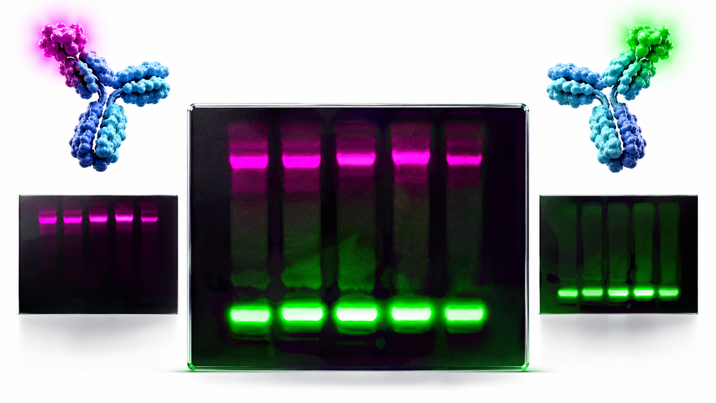
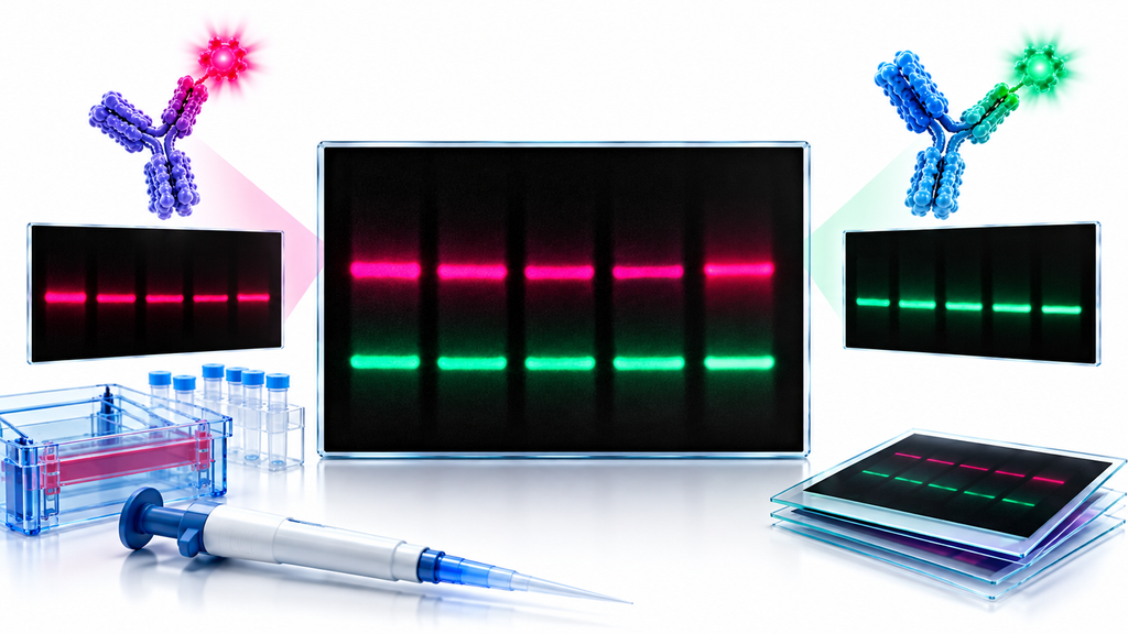
When the target protein and loading control run at sufficiently different molecular weights, both can be detected simultaneously using different fluorescence channels (Cy3/IR700 and Cy5/IR800 on LI-COR Odyssey or similar systems) or by using near-infrared secondary antibodies with low spectral overlap.
Target range
Recommended loading control
Detection strategy
Above 60 kDa
Alpha-tubulin (55 kDa)
ECL: sequential; fluorescence: multiplex
30 to 55 kDa
ACTB (42 kDa) or GAPDH (37 kDa)
Choose control furthest from target MW; confirm no overlap
Below 30 kDa (e.g. small proteins, phosphopeptides)
GAPDH (37 kDa)
Run control near top of relevant size range
Nuclear fraction
Histone H3 (17 kDa)
Total H3 on separate identical blot when detecting H3 modifications
When to strip and reprobe
Strip the membrane when the loading control and target protein run at the same or overlapping molecular weights and multiplex detection is not available.
Use mild stripping buffer (glycine pH 2.0 or commercial mild stripping solution) to preserve antibody epitopes for the next round of detection. Harsh stripping buffers containing beta-mercaptoethanol can strip protein from the membrane and reduce signal in subsequent probings.
After stripping, confirm the membrane is clear by exposing to ECL or imaging by fluorescence before applying the next primary antibody.
Validating Your Loading Control Before Use
Not all housekeeping antibodies are equal. A loading control antibody must meet three criteria before being used for quantification: it must produce a single clean band at the expected molecular weight, band intensity must increase linearly with loaded protein amount, and expression must remain stable under the experimental conditions being tested.
Validation step
How to perform it
What to look for
Single band confirmation
Run on standard cell lysate panel; image with ECL
One band at expected MW; no secondary bands above 15% of primary band intensity
Linear range test
Load 5, 10, 20, 40 µg protein in serial dilutions; plot band intensity vs. load
R-squared above 0.98 across the loading range used in experiments
Treatment stability
Run control in treated vs. untreated matched samples before full experiment
Less than 10% variation in normalized band intensity across treatment groups
Lot-to-lot consistency (recombinant antibodies)
Test new lot in parallel with current lot on identical samples
Band pattern and intensity within 15% of previous lot
AbTrivia recombinant monoclonal antibodies are produced from defined gene sequences, giving consistent lot-to-lot performance that simplifies this validation step — the linear range and band pattern established with one lot apply to all subsequent lots of the same clone.
Frequently Asked Questions
What is a loading control in Western blot
A loading control is a housekeeping protein — most commonly ACTB, GAPDH, tubulin, or Histone H3 — used to confirm equal protein loading across lanes and to normalize target band intensity for quantification.
Can I use GAPDH as a loading control for all experiments
No, because GAPDH expression is altered by hypoxia and glycolysis-modifying treatments, and it is absent from nuclear fractions — validate GAPDH stability under your specific experimental conditions before using it for normalization.
Which loading control should I use for nuclear extracts
Histone H3 is the only standard loading control appropriate for nuclear fractions, because ACTB, GAPDH, and tubulin are cytoplasmic proteins not reliably present in pure nuclear lysate.
My target protein and ACTB run at similar molecular weights — what should I use
Switch to alpha-tubulin (55 kDa) if your target is above 55 kDa, or to GAPDH (37 kDa) if your target is above 42 kDa, choosing whichever gives the greatest separation from the target band.
Should I strip and reprobe for the loading control or run it simultaneously
Run it simultaneously using multiplex fluorescence detection when possible, because stripping introduces additional protocol steps and risks protein loss from the membrane.
How do I normalize my Western blot data using a loading control
Divide the target protein band density by the loading control band density from the same lane for each sample, then calculate fold change using these normalized values relative to the control condition.
Do I need a loading control if I use equal protein concentration by BCA
Yes, because BCA confirms protein concentration in solution but does not account for gel loading variation, transfer efficiency differences, or pipetting error during sample preparation.
Can I use the same loading control antibody for all cell lines
Validate expression levels in each cell type before assuming stability, because some cell lines and primary cell types show altered housekeeping protein expression that makes standard controls unreliable for quantification.
Final Thoughts
Choosing the right loading control is not a default decision — it depends on sample type, target protein molecular weight, and experimental conditions.
ACTB and GAPDH work for standard cell lysates and most tissue samples but are inappropriate for muscle tissue, nuclear fractions, and experiments that directly alter actin or glycolytic pathways. Alpha-tubulin is the best choice for high-molecular-weight targets. Histone H3 is required for nuclear fraction normalization and chromatin studies.
AbTrivia supplies validated loading control antibodies for all four major housekeeping proteins — ACTB, GAPDH, tubulin, and Histone H3 — across monoclonal, polyclonal, and recombinant monoclonal formats, each with confirmed single-band data, linear range validation, and multi-species reactivity documented on every product datasheet.
Fast & Free Shipping
Free shipping on orders $999+
Quality Guarantee
Advenit post multos Scudilo Scutariorum tribunus velament
Secure Payment
We accept credit cards, PayPal, and bank wires
Lifetime Customer Support
Responsive and knowledgable customer service via email, text or phone