Hybridoma vs Phage Display vs Single B Cell: Antibody Discovery Compared
Choosing between hybridoma vs phage display vs single B cell methods can shape how quickly you find an antibody and how well it fits your research. Hybridoma technology creates antibody-producing cell lines from immunized animals, while phage display antibody discovery screens large libraries for target binders. Single B cell antibody screening recovers antibodies directly from individual immune cells, often preserving their natural heavy- and light-chain pairing. Each approach offers different benefits for antibody diversity, affinity, cost, and development time. This guide compares the three antibody discovery platforms, explains their workflows, and helps you select the right method for your project.
What Are the Three Main Antibody Discovery Platforms?
Antibody discovery platforms identify antibodies that recognize specific antigens. The three established approaches are hybridoma technology, phage display, and single B cell screening.
These monoclonal antibody discovery methods differ in how antibody candidates are generated, selected, and produced. Each can support research, diagnostic development, and therapeutic antibody discovery.
Hybridoma Technology
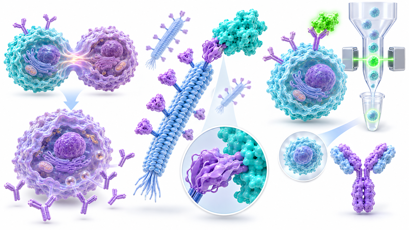
Hybridoma technology combines antibody-producing B cells with immortal myeloma cells. The resulting hybridoma can continuously produce a monoclonal antibody, making this approach useful for research reagents and diagnostic development.
Phage Display Technology
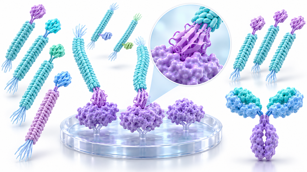
Phage display antibody discovery uses engineered bacteriophages to display antibody fragments. Researchers screen large libraries against a target antigen and recover the genetic sequences of promising binders.
Single B Cell Technology
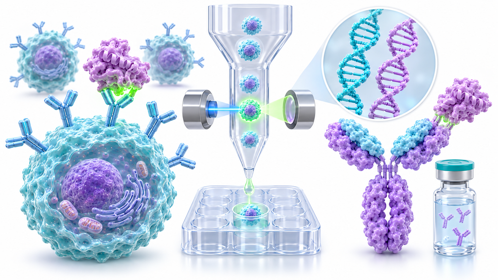
Single B cell antibody screening isolates individual antigen-specific B cells and recovers their antibody sequences. This method helps preserve natural heavy-light chain pairing while supporting recombinant antibody production.
The hybridoma antibody production method begins with immunizing an animal against a selected antigen. Researchers then isolate antibody-producing B cells and fuse them with myeloma cells.
This approach connects in vivo immunization with laboratory screening. Successful hybridoma clones can produce antibodies continuously, although clone stability and antibody specificity still require validation.
Step 1: Antigen Preparation and Immunization
Researchers prepare a suitable antigen and immunize an animal, commonly a mouse. Repeated immunization may improve the response and generate antigen-specific B cells with useful binding properties.
Step 2: B Cell Isolation and Fusion
Antibody-producing B lymphocytes are isolated from immune tissues and combined with myeloma cells through myeloma cell fusion. Successful fusion creates cells capable of antibody production and continued growth.
Step 3: Hybridoma Selection and Screening
Researchers use HAT selection medium to select hybridomas while eliminating unfused myeloma cells. Surviving cultures are screened for antigen binding, commonly through ELISA or related assays.
Step 4: Clone Selection and Antibody Production
Positive hybridoma clones undergo further cloning to establish monoclonal populations. Selected hybridoma cell lines are expanded, and their antibodies are purified and tested for the intended application.
Advantages and Limitations of Hybridoma Technology
Hybridomas provide established production workflows and preserve native heavy-light pairing in the original antibody-producing cell. However, fusion efficiency varies, some clones become unstable, and animal immunization requires additional time.
Phage display antibody discovery uses in vitro antibody selection instead of requiring animal immunization for every project. Antibody fragments are displayed on bacteriophages and screened against the desired antigen.
The method connects each displayed antibody with its genetic sequence. This allows researchers to identify promising binders and recover their sequences for further engineering.
Step 1: Create an Antibody Library
A phage display library contains many antibody variants, commonly displayed as single-chain variable fragments (scFv) or antigen-binding fragments (Fab).
Libraries may be naïve, immune-derived, or synthetic.
Step 2: Display Antibody Fragments on Phages
The bacteriophage M13 system commonly displays antibody fragments through a coat-protein fusion. This connects antibody binding properties with the DNA encoding the displayed fragment.
Step 3: Perform Biopanning Selection
During phage display biopanning, the library is exposed to the target antigen. Nonbinding phages are removed, while bound phages are recovered and amplified for additional screening.
Step 4: Identify and Validate Antibody Candidates
After biopanning selection rounds, researchers sequence enriched clones and evaluate their binding properties. Promising fragments may be converted into full-length antibodies for further functional testing.
Advantages and Limitations of Phage Display
Phage display supports large-scale antibody library screening and controlled selection conditions. However, conventional combinatorial libraries may disrupt native heavy-light pairing, and strong fragment binding doesn't guarantee successful full-length IgG performance.
A scientific review of phage display antibody libraries explains library design, selection strategies, and antibody developability.
Single B cell antibody screening identifies antibodies directly from individual immune cells. Researchers may use samples from immunized animals, vaccinated donors, or individuals with relevant immune responses.
Unlike many conventional phage libraries, this method can preserve native heavy-light chain pairing. It also allows direct recovery of antibody sequences without first creating immortalized hybridoma cells.
Step 1: Isolate Antigen-Specific B Cells
Researchers enrich antigen-specific B cells from blood or immune tissues. Fluorescently labeled antigens and fluorescence-activated cell sorting (FACS) can identify suitable memory B cells.
Step 2: Screen Individual B Cells
Microfluidic single-cell screening can capture secreted antibodies from individual cells. This helps identify rare antigen-specific clones and may support functional screening before sequence recovery.
Step 3: Recover Heavy and Light Chain Sequences
Single-cell RT-PCR or paired sequencing methods recover antibody variable regions. Maintaining the original VH and VL relationship is important for preserving the antibody's natural binding characteristics.
Step 4: Express and Validate Antibodies
Recovered sequences undergo antibody gene cloning and recombinant IgG expression. Researchers then test the resulting antibodies for binding specificity, affinity, purity, and functional activity.
Advantages and Limitations of Single B Cell Discovery
Single B cell technology offers direct sequence recovery and access to naturally selected antibodies. However, success depends on sample availability, sorting accuracy, and the frequency of suitable antigen-specific cells.
Hybridoma vs Phage Display vs Single B Cell: Key Differences
Hybridoma vs Phage Display vs Single B Cell methods differ in their starting materials, screening strategies, sequence recovery, and production requirements.
Hybridoma depends on cell fusion, phage display selects antibody fragments from libraries, and single B cell methods recover sequences directly from individual immune cells.
Antibody Discovery Platform Comparison
Feature
Hybridoma
Phage Display
Single B Cell
Main approach
B cell fusion
In vitro library selection
Individual B cell screening
Animal immunization
Usually required
Optional
Depends on source
Human antibody discovery
Requires suitable specialized methods
Supported by human libraries
Supported by human donor cells
Native VH/VL pairing
Preserved initially
Often rearranged in conventional libraries
Preserved with paired recovery
Sequence recovery
Requires additional work
Direct from selected clones
Direct from individual cells
Screening scale
Clone-based
Very large libraries
Depends on platform
Main limitation
Fusion efficiency and clone stability
Selection and display bias
Sample access and rare-cell recovery
Production
Hybridoma or recombinant
Recombinant
Recombinant
Which Platform Is Faster?
The antibody discovery timeline depends on antigen preparation, screening complexity, and validation requirements. Phage display and single B cell workflows may recover sequences sooner, while traditional hybridoma development requires cell fusion and clone stabilization.
Which Platform Costs Less?
An antibody discovery cost comparison should include immunization, sample access, equipment, labor, screening, sequencing, and production. Hybridoma may need extensive cell culture, while advanced single-cell methods can require specialized instruments.
There isn't one consistently cheapest platform.
Scientific and Technical Factors in Choosing an Antibody Discovery Platform
Choosing between Hybridoma vs Phage Display vs Single B Cell requires more than comparing screening speed. Antigen structure, antibody affinity, specificity, and downstream production requirements all influence success.
You should also consider antibody developability, especially when candidates are intended for therapeutic research. Strong antigen binding alone doesn't guarantee a suitable final antibody.
Antigen Presentation and Epitope Recognition
Antigen presentation affects which antibodies are selected. Purified proteins may expose different surfaces than native membrane proteins, so cell-based screening can be valuable for conformational epitopes.
Antibody Affinity and Binding Kinetics
Antibody binding kinetics describe how quickly an antibody binds and dissociates from its target. SPR and BLI can measure these interactions and help compare candidates beyond simple ELISA signals.
Specificity and Cross-Reactivity
Binding specificity must be tested against related proteins and relevant biological samples. An antibody may bind its intended antigen strongly while also recognizing unrelated structures.
Antibody affinity maturation can improve selected binders through targeted sequence changes and further screening. Phage display supports iterative library selection, while antibodies from other platforms can also undergo engineering.
Stability, Aggregation, and Expression
Antibody stability and aggregation affect storage, manufacturing, and experimental performance. Evaluate expression yield, solubility, thermal stability, and aggregation alongside affinity.
7. Which Antibody Discovery Platform Is Best for Your Application?
The best platform depends on your biological target and final application. Hybridoma remains useful for established monoclonal reagent development, while phage display supports extensive library selection.
Single B cell vs hybridoma decisions often depend on the importance of rapid sequence recovery, available immune cells, and preferred production methods.
For Research and Diagnostic Antibodies
Hybridoma technology works well when researchers need established clone-based production and application-specific screening. However, recombinant expression can also provide consistent supply after antibody sequencing.
Fully human antibody discovery may use human phage libraries or human donor B cells. Both can produce human-sequence candidates, but therapeutic suitability still depends on extensive safety and functional assessment.
For Therapeutic Antibody Development
Therapeutic antibody discovery requires more than binding affinity. Evaluate target function, epitope recognition, sequence liabilities, cross-reactivity, expression, stability, and potential immune effects before selecting a lead candidate.
For Difficult Antigens and Membrane Proteins
Membrane proteins may require native cell-based antigen presentation. Hybridoma and single B cell approaches can support such screening, while phage display can use cell-based selection strategies.
Application-specific hybridoma, phage, or single B cell screening
For established candidates, AbTriva's recombinant antibody collection offers examples of sequence-defined research reagents.
Frequently Asked Questions
1. What are the four types of monoclonal antibodies?
The four traditional types of therapeutic monoclonal antibodies are murine, chimeric, humanized, and fully human. Murine antibodies originate from mice, chimeric antibodies combine human and nonhuman regions, humanized antibodies contain mostly human sequences, and fully human antibodies use human antibody sequences.
2. How does phage display work for antibody discovery?
Phage display presents antibody fragments on the surface of bacteriophages. These phages are exposed to a target antigen, and nonbinding phages are washed away. Binding phages are recovered, amplified, and screened through repeated biopanning rounds to identify antibodies with desirable binding properties.
3. What is the key difference between hybridomas and recombinant antibodies?
Hybridoma technology produces monoclonal antibodies using immortalized antibody-secreting cells created by fusing B cells with myeloma cells. Recombinant antibodies are expressed from defined antibody gene sequences. Recombinant production generally offers better sequence control and batch consistency, while hybridoma-derived antibodies can also be converted into recombinant formats.
4. What is the difference between phage display and yeast surface display?
Phage display presents antibody fragments on bacteriophages and typically selects binders through biopanning. Yeast surface display presents antibodies or fragments on yeast cells and commonly uses fluorescence-activated cell sorting (FACS). Yeast enables quantitative binding analysis, while phage display generally supports larger antibody libraries.
5. What is the typical antibody discovery workflow?
The antibody discovery workflow begins with target identification and antigen preparation, followed by antibody generation using hybridoma, phage display, or single B cell technology. Researchers then screen promising candidates, sequence their antibody genes, perform recombinant expression, and evaluate binding affinity, specificity, functionality, and developability before selecting lead antibodies.
Conclusion
Hybridoma vs Phage Display vs Single B Cell technologies offer different routes to discovering monoclonal antibodies. Hybridoma provides established cell-based production, phage display enables extensive in vitro screening, and single B cell methods support direct recovery of naturally paired antibody sequences.
The best choice depends on antigen properties, research goals, available resources, and downstream applications. Carefully designed screening and functional validation remain essential regardless of the platform selected.
Fast & Free Shipping
Free shipping on orders $999+
Quality Guarantee
Advenit post multos Scudilo Scutariorum tribunus velament
Secure Payment
We accept credit cards, PayPal, and bank wires
Lifetime Customer Support
Responsive and knowledgable customer service via email, text or phone