• Contact info@abtriva.com for inquiries and orders.
  • Chinese (Simplified)

  • English

  • German

  • Korean

  • Spanish

United States (English / $ USD)

Rabbit anti-Human coronavirus 229E S Polyclonal Antibody

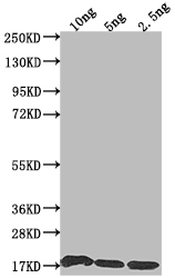
The antibody against S was raised in rabbit using the Recombinant Human coronavirus 229E Spike glycoprotein(S),partial (785-873aa) as the immunogen. This antibody exists as a non-conjugated isotype IgG, Protein G Purified. This antibody has been validated on ELISA, WB.

ADC-54714A

The antibody against S was raised in rabbit using the Recombinant Human coronavirus 229E Spike glycoprotein(S),partial (785-873aa) as the immunogen. This antibody exists as a non-conjugated isotype IgG, Protein G Purified. This antibody has been validated on ELISA, WB.

$299.00

Number
Order Exclusive Products Now

Request a Quote
High Purity LevelsPrecision and ReliabilityCustomization Options

Specifications


Cat.No ADC-54714A ClonalityPolyclonal
Host SpeciesRabbitTarget NameS
Target SynonymsS, Spike glycoprotein (S glycoprotein) (E2) (Peplomer protein)FormLiquid
Species ReactivityHuman coronavirus 229EIsotypeIgG
Storage Buffer0.01M PBS, 0.03% Proclin 300; Constituents: 50% Glycerol, PH 7.4Purification MethodProtein G purified
ConjugateNon-conjugatedApplicationELISA, WB
StorageUpon receipt

Immunogen Information


Immunogen DescriptionRecombinant Human coronavirus 229E Spike glycoprotein(S),partial (785-873aa)Target SpeciesHuman coronavirus 229E
Immunogen SequenceComplete sequences for the immunogen, target protein, and peptides are available upon request.Uniprot IDP15423
Background Information
  • Uniprot Id

    P15423

  • Target Species

    Human coronavirus 229E

  • Target Name

    S

  • Target Function

    S1 region attaches the virion to the cell membrane by interacting with host ANPEP/aminopeptidase N, initiating the infection. Binding to the receptor probably induces conformational changes in the S glycoprotein unmasking the fusion peptide of S2 region and activating membranes fusion. S2 region belongs to the class I viral fusion protein. Under the current model, the protein has at least 3 conformational states: pre-fusion native state, pre-hairpin intermediate state, and post-fusion hairpin state. During viral and target cell membrane fusion, the coiled coil regions (heptad repeats) regions assume a trimer-of-hairpins structure, positioning the fusion peptide in close proximity to the C-terminal region of the ectodomain. The formation of this structure appears to drive apposition and subsequent fusion of viral and target cell membranes.

  • Target Subcellular Location

    Virion membrane; Single-pass type I membrane protein. Host endoplasmic reticulum-Golgi intermediate compartment membrane; Single-pass type I membrane protein.

  • Target Protein Families

    Alphacoronaviruses spike protein family

  • Target Background

    S1 region attaches the virion to the cell membrane by interacting with host ANPEP/aminopeptidase N, initiating the infection. Binding to the receptor probably induces conformational changes in the S glycoprotein unmasking the fusion peptide of S2 region and activating membranes fusion. S2 region belongs to the class I viral fusion protein. Under the current model, the protein has at least 3 conformational states: pre-fusion native state, pre-hairpin intermediate state, and post-fusion hairpin state. During viral and target cell membrane fusion, the coiled coil regions (heptad repeats regions assume a trimer-of-hairpins structure, positioning the fusion peptide in close proximity to the C-terminal region of the ectodomain. The formation of this structure appears to drive apposition and subsequent fusion of viral and target cell membranes.

Inquire Rabbit anti-Human coronavirus 229E S Polyclonal Antibody Now


AbTriva respects your privacy and protects your personal data in accordance with AbTriva. For more information, please see our data protection statement. *

Notification