Flow Cytometry Staining Protocol: Surface and Intracellular Markers
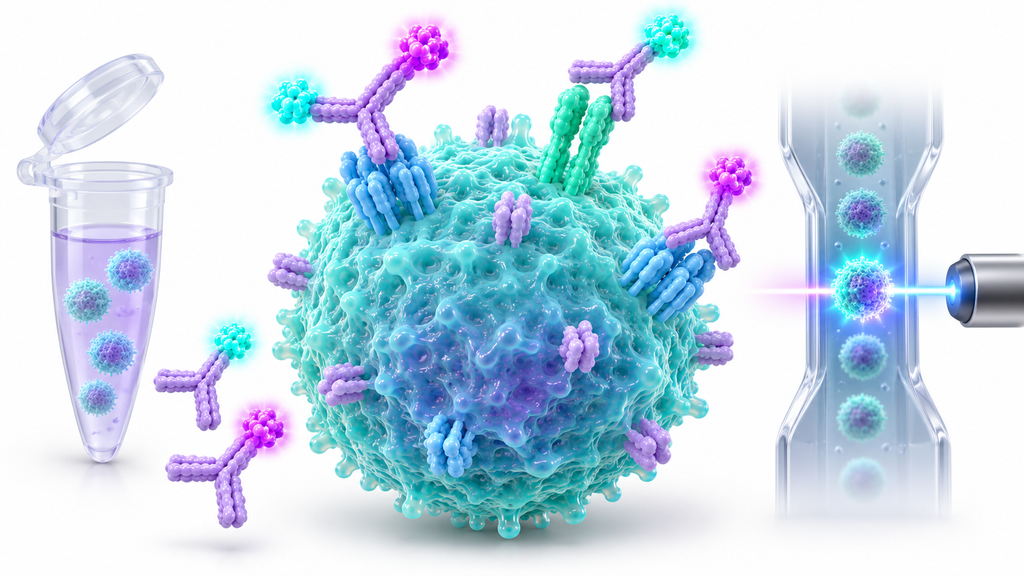
Flow cytometry helps researchers identify and characterize cells by detecting proteins on their surfaces and inside them. A reliable flow cytometry staining protocol begins with healthy cells, suitable antibodies, and proper sample preparation. Cell surface staining detects membrane proteins such as CD3 and CD4, while intracellular staining identifies cytokines, transcription factors, and signaling proteins. The main difference is that intracellular detection generally requires fixation and permeabilization to make internal targets accessible. Antibody selection, fluorochrome compatibility, and experimental controls also affect staining quality. This guide explains both methods, their step-by-step procedures, essential reagents, common problems, and practical ways to obtain reproducible results.
Surface vs. Intracellular Flow Cytometry Staining: Key Differences
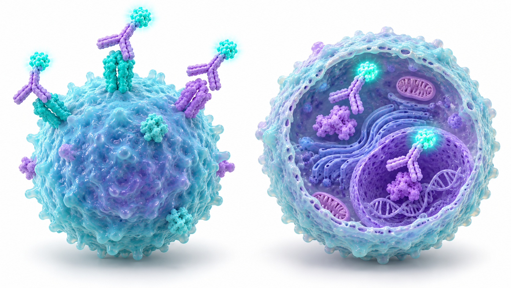
Surface vs. intracellular staining depends on where the target protein is located. Surface staining detects accessible membrane proteins, while intracellular staining identifies proteins within the cytoplasm or nucleus.
Surface staining usually works with intact cells. Intracellular staining generally requires fixation and permeabilization, which changes how cells and antibodies must be handled.
What Is Cell Surface Staining?
Cell surface staining uses fluorochrome-conjugated antibodies to detect proteins such as CD3, CD4, CD8, and CD19. Researchers use these markers to identify immune cell populations and examine changes in surface protein expression.
What Is Intracellular Staining?
Intracellular staining identifies proteins such as IFN-γ, TNF-α, and FOXP3. Fixation preserves cellular structures, while permeabilization allows antibodies to enter cells. The required buffer depends on whether the target is cytoplasmic or nuclear.
Surface vs. Intracellular Staining Comparison
Feature
Surface Staining
Intracellular Staining
Target location
Cell membrane
Cytoplasm or nucleus
Common markers
CD3, CD4, CD8, CD19
IFN-γ, TNF-α, FOXP3
Fixation
Often optional
Generally required
Permeabilization
Usually unnecessary
Required
Live-cell analysis
Possible
Not possible after fixation
Main challenge
Nonspecific surface binding
Epitope and buffer compatibility
Application
Immunophenotyping
Cytokine and signaling analysis
Both methods require antibodies validated for the intended sample and application. AbTriva's research antibody collection includes different antibody formats for laboratory studies.
Materials and Reagents for Flow Cytometry Staining
A successful flow cytometry staining protocol requires suitable cell suspensions, staining buffers, antibodies, and detection equipment.
The exact materials depend on the target protein, cell type, and whether fixation is necessary. Prepare compatible reagents before beginning the experiment.
Essential Buffers and Reagents
Common materials include PBS, bovine serum albumin (BSA), fluorochrome-conjugated antibodies, viability dyes, and Fc blocking reagents. Intracellular staining additionally requires fixation and permeabilization buffers selected for the target protein.
Choosing Suitable Antibodies
Choose antibodies validated for flow cytometry and your sample species. Review clone specificity, fluorochrome compatibility, and recommended dilution before use.
Healthy, well-separated cells produce more reliable staining. Dead cells may bind antibodies nonspecifically, while clumps interfere with individual cell measurements. Gentle handling and appropriate filtration help maintain a suitable single-cell suspension.
Cell Surface Staining Protocol for Flow Cytometry
A cell surface staining protocol measures proteins exposed on intact cell membranes. The workflow includes sample preparation, viability assessment, Fc receptor blocking, antibody incubation, and washing.
Exact conditions vary by cell type and reagent. Use validated recommendations when selecting antibody concentrations, incubation temperatures, and wash conditions.
Step 1: Prepare the Single-Cell Suspension
Collect cells and resuspend them in a compatible staining buffer. Gently remove aggregates or debris when necessary.
Maintain consistent cell numbers between samples, since cell concentration affects antibody availability and staining performance.
Step 2: Perform Cell Viability Staining
Cell viability staining identifies dead cells that may contribute to nonspecific fluorescence. Use a fixable viability dye when subsequent fixation is planned.
For unfixed samples, membrane-impermeant dyes such as PI or 7-AAD may be appropriate.
Step 3: Block Fc Receptors
Fc receptor blocking reduces unwanted antibody binding to Fc receptor-expressing cells, including monocytes and macrophages.
Mouse experiments commonly use CD16/CD32 blocking reagents. Human samples require suitable human Fc receptor blocking reagents.
Add antibodies at validated concentrations and mix gently. Many surface staining procedures use approximately 20–30 minutes at 4°C, protected from light.
For defined marker detection, review suitable monoclonal antibodies and confirm flow cytometry validation.
Step 5: Wash and Prepare Cells for Acquisition
Wash cells to remove unbound antibodies, then resuspend them in the appropriate acquisition buffer.
Analyze samples using compatible instrument settings. Avoid unnecessary delays, as changes in cell quality can affect fluorescence measurements.
Intracellular Staining Protocol for Flow Cytometry
A flow cytometry staining protocol for intracellular targets requires antibody access to proteins inside cells. Fixation and permeabilization are therefore central preparation steps.
Cytokines, transcription factors, and phosphoproteins have different requirements. Selecting compatible reagents helps preserve target recognition and fluorescence signals.
Step 1: Prepare Cells for Intracellular Antigen Detection
Cytokine studies may require cell stimulation before staining. PMA/ionomycin or receptor-specific stimulation can induce selected cytokines.
Brefeldin A or monensin may help retain intracellular cytokines. Conditions depend on the target and cell type.
Step 2: Perform Surface Staining Before Fixation
When combining surface and intracellular targets, stain compatible surface markers before fixation.
This helps preserve fixation-sensitive epitopes. However, some phosphoprotein experiments require immediate fixation to capture rapidly changing signaling states.
Step 3: Fix and Permeabilize the Cells
Paraformaldehyde-based fixation is commonly used to preserve cellular proteins. Permeabilization then allows antibodies to reach intracellular targets.
Some phosphoprotein assays require methanol treatment. Check compatibility with fluorochromes and phospho-specific antibodies.
Step 4: Stain Intracellular Cytokines and Transcription Factors
Cytokine staining commonly measures IFN-γ, TNF-α, and IL-2.
Nuclear proteins such as FOXP3, T-bet, and GATA3 usually require specialized transcription factor staining buffers. Maintain compatible permeabilization conditions during antibody incubation and washing.
Step 5: Wash and Analyze the Stained Cells
Wash using the buffer recommended for your fixation and permeabilization system.
Protect cells from light and resuspend them in a compatible acquisition buffer. Apply consistent instrument settings when comparing samples.
Choosing the Right Intracellular Staining Method
Target Type
Examples
Preparation Approach
Cytoplasmic cytokines
IFN-γ, TNF-α, IL-2
Compatible intracellular fixation and permeabilization
Nuclear proteins
FOXP3, T-bet, GATA3
Specialized nuclear staining buffer
Phosphoproteins
p-STAT3, p-ERK, p-AKT
Validated fixation and permeabilization
Surface and intracellular targets
CD4 with IFN-γ
Combined staining with compatible reagents
How to Choose Fluorochromes and Titrate Antibodies
Fluorochrome selection affects marker separation, signal brightness, and spectral interference.
Choose fluorescent dyes according to your cytometer's lasers, detectors, and target expression levels. Antibody titration helps determine suitable concentrations without excessive background.
Fluorochrome Selection for Flow Cytometry
Common fluorochromes include FITC, PE, APC, PerCP, and Alexa Fluor dyes.
Bright fluorochromes often suit low-expression markers, while abundant targets may tolerate dimmer dyes. Confirm compatibility with fixation and permeabilization.
Understanding Spectral Overlap and Compensation
Spectral overlap occurs when fluorescence from one dye contributes to another detector.
Conventional compensation or spectral unmixing separates overlapping signals. Accurate analysis requires suitable single-stained controls and consistent instrument settings.
Why Antibody Titration Is Important
Antibody titration identifies a concentration that separates positive and negative populations clearly.
Test several concentrations using the intended sample. The staining index can support comparisons, while excessive antibody may increase background.
Direct vs. Indirect Antibody Staining
Direct staining uses fluorochrome-conjugated primary antibodies. Indirect staining uses an unlabeled primary followed by a fluorescent secondary antibody.
Essential Controls for Surface and Intracellular Flow Cytometry
Reliable flow cytometry controls help distinguish genuine marker expression from autofluorescence, spectral interference, and nonspecific antibody binding.
A dependable flow cytometry staining protocol includes controls suited to the panel, sample preparation, and biological question.
Unstained and Single-Stained Controls
Unstained cells reveal baseline autofluorescence.
Single-stained controls provide reference signals for compensation or spectral unmixing. Use fluorochromes and preparation conditions compatible with the experimental samples.
Fluorescence Minus One (FMO) Controls
Fluorescence Minus One (FMO) controls contain every fluorochrome except the marker being evaluated.
They help establish gating boundaries, particularly when populations overlap or fluorescence spreading makes weak signals difficult to identify.
Isotype Controls and Fc Blocking
Isotype controls may help investigate some forms of nonspecific antibody binding, but they don't replace FMO controls.
Fc blocking is particularly important for cells expressing Fc receptors, since unwanted antibody attachment can increase background.
Positive and Negative Biological Controls
Positive controls confirm that the target can be detected, while negative controls help evaluate specificity.
Begin by excluding debris and doublets. Next, use a viability gate to remove dead cells before identifying relevant populations.
Common Flow Cytometry Staining Problems and Troubleshooting
Flow cytometry troubleshooting starts with checking sample quality, antibody concentration, reagent compatibility, and instrument settings.
Weak fluorescence, high background, and poor cell recovery may have different causes. Investigate the affected stage before changing multiple experimental conditions.
Weak or Missing Fluorescence Signals
Weak signals may reflect low antigen expression, insufficient antibody, poor fluorochrome selection, or incompatible fixation.
For intracellular targets, confirm that permeabilization provides antibody access without damaging the relevant epitope.
High Background and Nonspecific Antibody Binding
High background may result from excess antibody, dead cells, inadequate washing, or Fc receptor interactions.
Repeated washing, harsh centrifugation, and clumping can reduce cell recovery.
Fixation may also change forward- and side-scatter patterns. Review cell quality and gating before interpreting unexpected populations.
Flow Cytometry Troubleshooting Table
Problem
Possible Cause
Recommended Action
Weak fluorescence
Low antigen expression or insufficient antibody
Optimize staining concentration and fluorochrome
High background
Dead cells or nonspecific binding
Improve viability gating and Fc blocking
Poor population separation
Spectral spreading or unsuitable dye
Review fluorochrome selection and FMO controls
Cell clumping
Inadequate sample preparation
Use gentle mixing and suitable filtration
Low cell recovery
Harsh washing or centrifugation
Review handling conditions
Unexpected intracellular signal
Incompatible fixation or permeabilization
Verify buffer and antibody compatibility
Best Practices for Reliable Surface and Intracellular Staining
A reliable flow cytometry staining protocol begins with selecting the correct staining approach for your target.
Healthy samples, validated antibodies, compatible fluorescent labels, and appropriate controls support consistent results. Intracellular targets require additional attention to fixation and permeabilization.
Choose the Right Protocol for Your Target Protein
Use surface staining for membrane proteins and intracellular staining for cytoplasmic or nuclear targets.
Phosphoproteins may require rapid fixation, while cytokines often need stimulation and secretion-blocking reagents. Match preparation conditions to target biology.
How to Combine Surface and Intracellular Staining
For many combined experiments, perform fixable viability staining and surface staining before fixation.
Then permeabilize, stain intracellular targets, wash, and acquire data. Confirm that the chosen antibodies remain compatible throughout the workflow.
Improve Antibody Selection and Sample Quality
Good flow cytometry panel design uses suitable fluorochromes, validated clones, and healthy cells.
AbTriva's recombinant antibody collection includes sequence-defined antibodies that researchers can evaluate for suitable applications after checking product-specific validation.
Frequently Asked Questions
Surface and intracellular staining answer different biological questions, but both rely on careful antibody selection and sample preparation.
These common questions address staining order, fixation, permeabilization, and controls.
What Is the Difference Between Surface and Intracellular Staining?
Surface staining detects proteins exposed on intact cell membranes. Intracellular staining measures proteins within the cytoplasm or nucleus and generally requires fixation and permeabilization to allow antibody access.
Can You Perform Surface and Intracellular Staining Together?
Yes. Both methods can be combined when antibodies, fluorochromes, and fixation conditions are compatible. Surface staining is commonly performed first, followed by fixation, permeabilization, and intracellular staining.
Should Surface Staining Be Done Before Fixation?
Usually, surface staining is completed before fixation when combined with intracellular markers. However, certain signaling studies require immediate fixation to preserve phosphorylation states. Follow a validated protocol for your target.
What Is the Best Permeabilization Buffer for Intracellular Flow Cytometry?
The best buffer depends on the target protein. Cytokines often use cytoplasmic permeabilization systems, while transcription factors require suitable nuclear buffers. Some phosphoproteins need methanol-based treatment.
What Controls Are Needed for Intracellular Flow Cytometry?
Common controls include unstained cells, single-stained references, viability controls, and appropriate positive and negative biological samples. FMO controls help establish gates, while isotype controls may investigate selected nonspecific binding effects.
Conclusion
Choosing the right flow cytometry staining protocol helps researchers detect surface and intracellular markers with greater confidence. Surface staining identifies membrane proteins, while intracellular staining requires suitable fixation and permeabilization.
For reliable results, prioritize sample quality, antibody specificity, fluorochrome compatibility, and experimental controls. Titrate antibodies, verify staining conditions, and use consistent acquisition settings. These practices improve population separation, reduce unwanted background, and support reproducible flow cytometry data.
Fast & Free Shipping
Free shipping on orders $999+
Quality Guarantee
Advenit post multos Scudilo Scutariorum tribunus velament
Secure Payment
We accept credit cards, PayPal, and bank wires
Lifetime Customer Support
Responsive and knowledgable customer service via email, text or phone