ELISA Troubleshooting: Weak Signal, High Background, and Poor Standard Curve
ELISA troubleshooting is most productive when the problem is matched to its root cause before any reagent or protocol change is made.
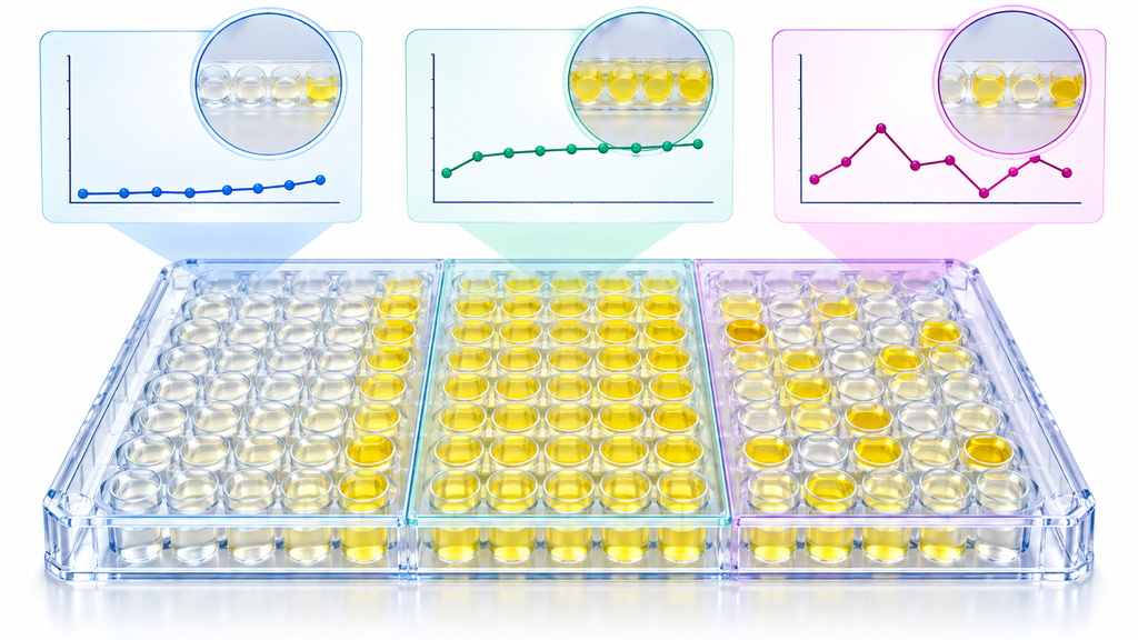
The three most common ELISA failures — weak or absent signal, high background absorbance, and a poor or flat standard curve — each have a distinct set of causes, and the fastest path to a working assay is identifying which category applies before changing anything.
This article covers each failure mode with diagnostic questions, ranked causes, and the most effective fixes for both sandwich ELISA and direct or indirect ELISA formats.
ELISA Formats and Why They Fail Differently
Sandwich ELISA uses a capture antibody coated to the plate, a sample containing the antigen, a detection antibody that binds a different epitope, and a signal-generating enzyme conjugate.
Direct and indirect ELISA detect antigen or antibody without a capture step. The failure modes overlap with sandwich ELISA but differ in which step is most likely responsible for each problem.
Format
Most common failure
First thing to check
Sandwich ELISA
Weak signal or flat standard curve
Capture antibody coating concentration and antigen-antibody pair compatibility
Indirect ELISA (antibody detection)
High background
Blocking agent and secondary antibody dilution
Direct ELISA (antigen detection)
Weak signal or no signal
Coating efficiency and antigen stability
Competitive ELISA
Poor standard curve shape
Standard concentrations, competitor antigen purity, and incubation order
Problem 1: Weak or Absent Signal
Weak signal means the absorbance readings from standard curve samples are lower than expected, sample readings fall below the detection limit, or the signal-to-blank ratio is less than 2. Absent signal means all wells including the high-concentration standards read at or near blank.
Diagnostic question: Did the positive control work
Always include a positive control — a known high-concentration sample or recombinant antigen — in every ELISA run. If the positive control also shows weak or absent signal, the problem is in the assay itself, not the samples.
If the positive control reads correctly and samples are weak, the issue is likely low analyte concentration in samples or a matrix interference problem.
Cause 1: Antibody not recognizing the antigen in the sample matrix
The capture or detection antibody may be validated against recombinant protein but not against the native antigen form present in the sample, particularly in serum, plasma, or tissue lysate where the protein may be glycosylated, truncated, or complexed with binding partners.
Confirm the antibody datasheet lists validation specifically in the sample type being tested. AbTrivia monoclonal antibodies and polyclonal antibodies for ELISA each include confirmed sample type reactivity and recommended assay formats on the product page.
Cause 2: Capture antibody coating inefficiency
Insufficient antigen capture is a primary cause of weak signal in sandwich ELISA. The capture antibody must coat the plate at adequate density to capture enough antigen from the sample for the detection antibody to generate signal.
Parameter
Typical starting condition
If signal is weak, try
Capture antibody concentration
1 to 4 µg/mL in carbonate buffer pH 9.6
Increase to 4 to 8 µg/mL; optimize pH
Coating volume
100 µL per well
Confirm consistent volume delivery across all wells
Coating incubation
Overnight at 4°C
Extend to 16 to 18 h; confirm plate was sealed during incubation
Coating buffer
PBS pH 7.4 or carbonate pH 9.6
Try the alternative buffer — some antibodies coat more efficiently at higher pH
Plate type
Standard 96-well ELISA plate
Switch to high-binding plate if using medium-binding; confirm plate is protein-binding grade
Cause 3: Detection antibody or conjugate problem
An incorrect secondary antibody, a degraded HRP conjugate, or an enzyme substrate prepared incorrectly are common causes of weak or absent signal that pass unnoticed because the coating and blocking steps appear normal.
Confirm the secondary antibody host and target species match the detection antibody
Check HRP conjugate storage conditions — HRP is inactivated by repeated freeze-thaw cycles and by sodium azide, which must be excluded from HRP-containing reagents
Prepare TMB substrate fresh from concentrated stock immediately before use; do not reuse substrate from a previous run
Verify stop solution concentration (typically 1 M H2SO4 or 2 N H2SO4) and confirm color changes from blue to yellow after adding stop solution
AbTrivia secondary antibodies for ELISA are available as HRP conjugates for anti-rabbit, anti-mouse, and anti-human primaries, with lot-tested activity confirmation documented on each product datasheet.
Cause 4: Sample matrix interference
Serum and plasma contain proteins, lipids, and other components that can reduce signal by competing with the analyte for binding sites, inhibiting the enzyme conjugate, or causing non-specific quenching of the colorimetric reaction.
Test a 1:2 and 1:4 dilution of the sample in assay diluent alongside the neat sample. If the diluted samples fall on the standard curve while the neat sample gives lower-than-expected signal, matrix interference is confirmed.
Apply a sample dilution factor of at least 1:2 in assay diluent as the standard starting condition for serum and plasma samples in all ELISA protocols.
Cause 5: Antigen degradation
Samples stored at minus 20°C for extended periods, subjected to repeated freeze-thaw cycles, or handled at room temperature for prolonged periods before assay can show reduced signal due to analyte degradation rather than assay failure.
Include protease inhibitor cocktail in sample preparation buffers for tissue lysates. Aliquot samples before freezing to prevent repeated freeze-thaw, and confirm sample integrity on a parallel Western blot before concluding that the ELISA signal is genuinely low.
Problem 2: High Background in ELISA
High background means the blank wells (no antigen, no sample) show elevated absorbance above 0.1 to 0.15, or the signal-to-blank ratio for the lowest standard is less than 2. High background raises the assay detection limit and makes low-concentration samples unquantifiable.
Cause 1: Insufficient blocking
Blocking fills non-specific protein-binding sites on the plate surface that would otherwise bind detection antibody or conjugate non-specifically and generate signal in all wells including blanks.
Blocking agent
Best for
Avoid when
1 to 3% BSA in PBS
Most ELISA formats; phospho-specific antigens
Biotinylated antibody detection systems (BSA contains biotin)
5% non-fat milk in PBST
Standard sandwich ELISA, most cytokine assays
Phospho-specific or phosphoprotein detection (milk contains casein, a phosphoprotein)
5% normal serum
Reducing non-specific binding in indirect ELISA
Use serum from secondary antibody species; not primary species
Commercial plate blocker
High-background problems resistant to standard blocking agents
0.5% casein in PBS
General use; low background alternative to milk
Phospho-specific detection
Extend blocking time from 1 hour to 2 hours if standard blocking conditions do not resolve background. Add 0.05% Tween-20 to the blocking buffer to improve non-specific binding suppression on high-protein samples.
Cause 2: Insufficient washing
Residual unbound antibody or conjugate left in wells from incomplete washing generates background signal across all wells after substrate addition.
Increase wash cycles from 3 to 5 per step. Ensure complete aspiration at each wash — wells that retain liquid carry over residual reagents into the next step. Allow 30 to 60 seconds of soaking time per wash cycle rather than immediately aspirating.
Cause 3: Detection antibody concentration too high
Excess detection antibody saturates the plate surface including non-specifically adsorbed molecules, generating signal above background even in wells with no antigen.
Titrate the detection antibody across a 4-fold dilution series — for example 0.25, 0.5, 1.0, and 2.0 µg/mL — and select the concentration that gives the highest signal-to-background ratio rather than the highest raw signal.
Cause 4: HRP conjugate non-specific binding
HRP-conjugated secondary antibodies or streptavidin-HRP conjugates can bind non-specifically to plate surfaces and blocking proteins if used at too high a concentration or with insufficient blocking.
Dilute the conjugate in blocking buffer (not in PBS alone), which provides carrier proteins that compete with non-specific plate binding. Standard starting concentrations for streptavidin-HRP are 1:1,000 to 1:10,000 from a 1 mg/mL stock.
Cause 5: Substrate incubation too long
TMB substrate generates signal as a time-dependent reaction. Extended substrate incubation increases background non-linearly, because even minimal non-specific HRP binding produces measurable signal given enough time.
Set a fixed substrate incubation time of 10 to 15 minutes and stop the reaction at that time regardless of color intensity. Do not extend incubation to increase low signals — address weak signal by optimizing upstream steps instead.
Problem 3: Poor Standard Curve
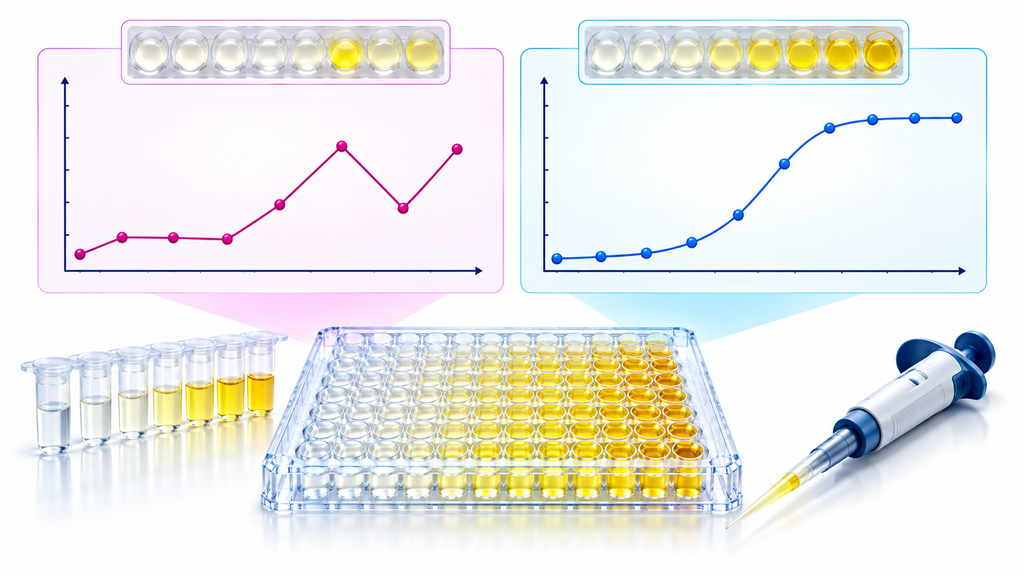
A standard curve problem means the curve is flat (all standards at similar OD), shows an irregular or non-sigmoidal shape when plotted, has poor R-squared on 4PL fitting, or the high-end standards are indistinguishable from each other (hook effect).
Cause 1: Standard preparation errors
Incorrect reconstitution of lyophilized standards, pipetting errors during serial dilution, or using an expired or degraded standard stock are the most common causes of flat or non-sigmoidal curves.
Standard problem
Most likely cause
Fix
All standards at same OD
Stock standard not reconstituted correctly; pipetting error in dilution
Reconstitute fresh standard; repeat serial dilution with calibrated pipettes
Top standard lower than expected
Standard degradation; incorrect concentration of stock
Use fresh stock; confirm reconstitution volume matches datasheet
Bottom standards all at blank level
Serial dilution error in lower wells; pipette carryover
Use fresh tips for each dilution transfer; confirm correct diluent volume in each well
Non-sigmoidal shape
Dilution factor incorrect for the assay range
Adjust concentration range — reduce top standard or increase bottom standard
Dilute samples and standards; reduce top standard concentration
Cause 2: Antibody pair incompatibility
In sandwich ELISA, the capture and detection antibodies must bind to two non-overlapping epitopes on the target antigen simultaneously. If the antibody pair recognizes overlapping or sterically interfering epitopes, the detection antibody cannot bind after the antigen is captured, producing a flat standard curve regardless of antigen concentration.
When using separately sourced capture and detection antibodies rather than a matched pair, confirm epitope mapping data on the datasheet or test an alternative detection antibody. AbTrivia polyclonal antibodies recognize multiple epitopes on the target protein and are often used as the detection antibody in sandwich pairs where the capture antibody is a monoclonal targeting a single defined epitope.
Cause 3: Wrong incubation conditions
ELISA antibody-antigen binding is temperature and time dependent. Incubations performed at the wrong temperature, for insufficient time, or without adequate mixing in the case of shake-plate formats can produce low signal across the entire standard curve.
Incubation step
Standard condition
Effect of deviation
Sample incubation (capture step)
1 to 2 h at RT with gentle shaking, or overnight at 4°C
Short incubation or no shaking reduces capture efficiency; overnight at 4°C improves sensitivity
Detection antibody incubation
1 h at RT with gentle shaking
Reduced time gives proportionally lower signal across all standards
HRP conjugate incubation
30 to 60 min at RT
Insufficient time reduces signal amplification
Substrate incubation
10 to 15 min at RT in the dark
Too short: flat curve; too long: saturation and hook effect at high concentrations
Cause 4: Plate reader or wavelength problem
Reading the plate at the wrong wavelength is a straightforward but overlooked cause of poor standard curve data. TMB read after stop solution requires 450 nm with a reference at 570 or 620 nm. TMB read kinetically (blue) before adding stop solution is read at 620 nm.
Confirm the plate reader wavelength setting matches the substrate and detection system being used. A mismatch produces uniformly low or erratic absorbance readings that do not reflect actual enzyme activity.
Proper controls distinguish assay failure from genuine biology and are required for publication-quality data.
Control
Purpose
How to include
Blank (diluent only)
Measures non-specific substrate signal and background
At least 2 wells per plate
Zero standard (diluent only in standard position)
Bottom anchor for 4PL curve fitting
Use assay diluent as the 0 pg/mL point
Positive control (known high-concentration sample)
Confirms assay is working across plates and runs
Same sample across all runs; track over time for QC
Matrix control (analyte-depleted sample matrix)
Detects matrix interference
Spike known concentration of standard into sample matrix
Inter-assay CV samples
Monitors plate-to-plate variability
Run in duplicate on every plate
AbTrivia tag and control antibodies include reagent-grade isotype controls and recombinant protein standards suitable for ELISA positive controls and spike-recovery experiments.
Phospho-Specific ELISA: Additional Considerations
Phospho-specific ELISA detects the phosphorylated form of a target protein and requires additional precautions that do not apply to standard total-protein assays.
Add phosphatase inhibitor cocktail to all lysis buffers and sample diluents to prevent dephosphorylation during sample preparation
Use BSA-based blocking only, never milk, because casein in milk is a phosphoprotein that directly competes with phospho-epitope detection
Keep samples on ice during all preparation steps and process quickly
Include a stimulated positive control (EGF-treated cells for p-EGFR, insulin-treated cells for p-AKT) and an unstimulated negative control on every plate
AbTrivia phospho-specific antibodies cover p-AKT S473, p-ERK1/2, p-STAT3 Y705, p-Smad3, and p-Histone H2AX, each with recommended ELISA conditions and blocking specifications documented on the product datasheet.
Frequently Asked Questions
Why does my ELISA give weak signal even when the antibody is validated
Validated for ELISA does not mean validated for your specific sample type, matrix, or species — confirm the datasheet lists confirmed reactivity for the exact sample preparation and species you are using.
What causes high OD in blank wells
Insufficient blocking, incomplete washing, or detection antibody concentration too high are the most common causes of elevated blank well absorbance.
Why is my standard curve flat
The most common causes are incorrect standard reconstitution or serial dilution, antibody pair epitope incompatibility, or incubation conditions that are too short or at the wrong temperature.
Can I reuse ELISA plates after washing
No, because coated capture antibody is stripped during washing and the plate cannot be reliably recoated for a second run.
How many washes are needed between ELISA steps
A minimum of 3 washes per step is standard, with 5 washes recommended when high background persists despite correct blocking conditions.
Why does my positive control read correctly but samples are weak
When the positive control signal is normal and samples are low, the most likely explanation is low analyte concentration in samples, matrix interference, or analyte degradation during sample storage.
What is the hook effect in ELISA
The hook effect occurs when antigen concentration exceeds the binding capacity of the capture antibody, causing the signal to plateau or drop at high concentrations and making the standard curve shape non-sigmoidal at the top.
Should I run standards in duplicate or triplicate
Run standards in duplicate as the minimum, with triplicate recommended for quantification data that will be used in publications or regulatory submissions.
Final Thoughts
ELISA troubleshooting is faster when the same diagnostic sequence is followed every time: confirm controls first, then check the failing step in isolation before changing multiple variables at once.
Most ELISA failures trace back to one of three sources — an antibody not suited to the sample type, a protocol step performed outside validated conditions, or reagent degradation that was not caught before the run began.
AbTrivia supplies ELISA-validated antibodies across all major formats, including monoclonal, polyclonal, recombinant monoclonal, and phospho-specific antibodies, each with confirmed ELISA application data, sample type reactivity, and recommended assay conditions on every product datasheet.
Fast & Free Shipping
Free shipping on orders $999+
Quality Guarantee
Advenit post multos Scudilo Scutariorum tribunus velament
Secure Payment
We accept credit cards, PayPal, and bank wires
Lifetime Customer Support
Responsive and knowledgable customer service via email, text or phone